Reading a Smile Like a Periodontist: The 5 Signs of a Truly Natural Implant Result
When a patient tells me a result looks natural, they're reacting to something they can't quite name. I'm checking five specific things when I look at the same case, and they all sit around the tooth, in the gum, the light, the way it moves, rather than in the tooth itself.
1. Papillary fill
The first thing I look at is the papilla, the small triangle of gum filling the space between two crowns. A restoration can be anatomically perfect and still read as artificial if that triangle is missing, blunted, or replaced by a dark gap where light reaches the palate. Papillary fill is slow biology. It depends on bone height, on the distance from contact point to crest, and on how carefully the soft tissue was handled at every stage of treatment. Surgery either preserves the papilla or it doesn't; no crown recovers it afterwards.
2. Scalloped symmetry
Natural gum tissue isn't flat. It follows a scalloped line that rises and falls with the root anatomy beneath it, with the gingival zenith, the highest point of the curve, sitting slightly distal to the long axis on the central incisors and canines. Repeat that curve consistently across the arch and the eye reads harmony before it registers why. Flatten it on even a single tooth, and something reads as off, even to someone who couldn't tell you what they're looking at.
3. The transition zone
This is the line I care about most, and the one almost nobody outside periodontics looks for directly: the point where the pink of the tissue meets the white of the restoration. In a well-managed case there's no visible margin, no grey shadow of metal, no cement line catching the light, no abrupt shift in colour. The tissue just continues into the tooth. I call this the Invisible Line, because done well, it disappears entirely — you stop being able to say where the biology ends and the prosthetics begin.
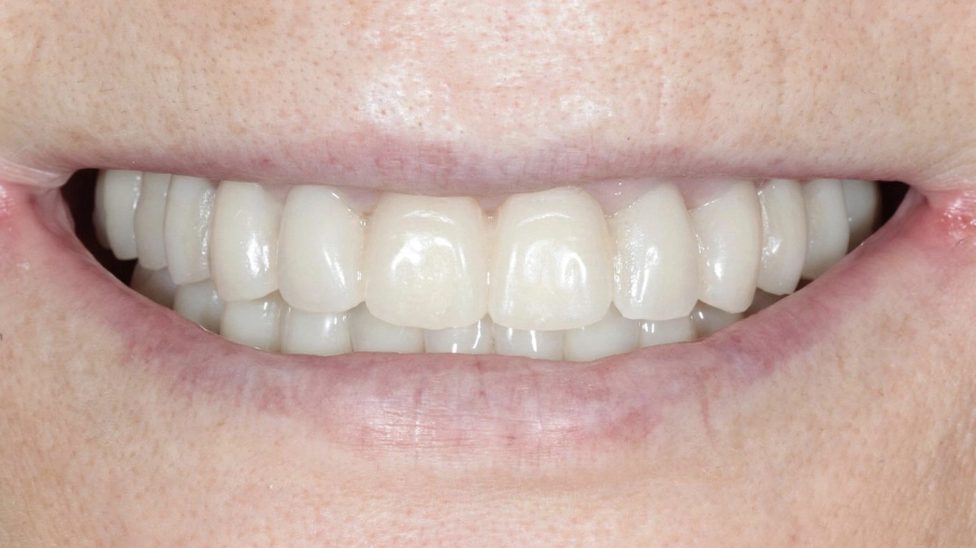
In this case, the papillae are present and full, the scallop is symmetrical left to right, and the transition zone holds, with the emergence profile carried through from the surgical stage to the final prosthetics without compromise. That's what lets the result sit inside the patient's own biology instead of on top of it.
4. Light behaviour
Natural enamel has translucency at the incisal third, subtle internal light scatter, and a surface texture (micro-ridges, perikymata) that breaks reflection into many small highlights instead of one flat shine. A restoration can photograph beautifully in a single frontal shot and still fail this test the moment the patient tilts their head under a different light, because it was built for the camera rather than for how light actually moves across enamel. I check this by watching patients move under different light sources before I call a case finished.
5. Function under animation
This one can't be judged from a static photograph, which is exactly why it gets skipped in marketing material. A result has to hold up while the patient is speaking, laughing, eating. Does the lip line still frame the restoration correctly through a full smile and at rest? Does the tissue blanch, or does the margin show under normal movement? If a result only works in a still frame, it was built for the camera rather than for the person wearing it.
Why this list is short
Five signs. Each one sits at an interface: bone and papilla, tissue and restoration, surgery and prosthetics, a static result and a functioning one. In implant dentistry, quality gets decided at the edges, in the zones most people are never trained to examine, rather than at the centre of the tooth where everyone happens to be looking.
The same principle applies well beyond a single restoration. I write more about it, and about what it means for organisations and clinical leadership, at The Invisible Line.
Full-Arch FP1 case documentation showing papillary fill, scalloped symmetry, and transition zone at follow-up.
View Case Documentation →