Full-Arch FP1 at Eight Years: Root Retention, Socket Shield and Connective Tissue Grafts in One Surgery
This is an eight-year follow-up of a maxillary full-arch FP1 rehabilitation. At the time of surgery the alveolar ridge was preserved by retaining root fragments (socket shield technique), connective tissue grafts were taken from the palate and from the maxillary tuberosity, and the maxillary canting was corrected, all in a single operation. At eight years there is no gingival recession and no loss of papillae. The only event in the follow-up is a small fracture of a lateral incisor on the ceramic prosthesis.
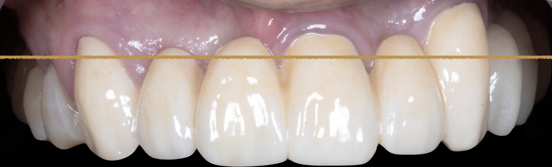
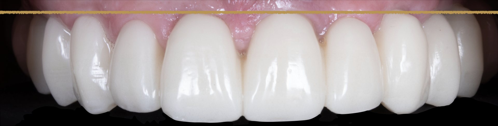
The first photograph shows the starting point, with uneven gingival margins and an incisal plane that is not level. The second shows the definitive prosthesis, eight years after surgery. The horizontal lines are references drawn on the photographs.
Why FP1 leaves no room for error
FP1 is the category of fixed prosthesis that replaces the crowns only, with no artificial gingiva. The result therefore depends on the patient’s own soft tissue. Height, thickness and papillae have to be right at the end of surgery, because there is no pink material to hide anything.
What was done in a single surgery
Four procedures were carried out in the same operation: partial extraction therapy with root retention, including the socket shield technique; connective tissue grafts from the palate and the tuberosity; correction of the maxillary canting; and implant placement with immediate provisional.
Root retention and the socket shield
After extraction the buccal bone plate remodels, and in the anterior maxilla this remodelling is a common cause of recession. Partial extraction therapy keeps part of the root and its periodontal ligament in place, which maintains the blood supply from the ligament to the surrounding tissues.
Connective tissue grafts from the palate and the tuberosity
Connective tissue grafts increase the thickness of the mucosa around implants. The graft can be harvested from the palate or from the tuberosity, and the two sites give tissue of different quality and with different morbidity. In this case I used both, taking advantage of their different properties.
Correction of the maxillary canting
Canting is a tilt of the maxillary plane relative to the horizontal. If it is left in place, the prosthesis either reproduces the tilt or hides it with unequal tooth lengths and pink material. With an FP1 neither option works, so the correction has to be part of the plan and of the surgery.
Doing these procedures in one operation meant a single healing period for the tissues, which were then shaped by the provisional prosthesis.
One year: the immediate provisional
The video shows the patient at one year with the fixed provisional in place. The provisional emerges from the gingiva like a natural tooth, the papillae fill the spaces between the teeth, and the soft tissue frames each tooth all the way round. With the provisional removed, the tissue keeps the contour that the prosthesis gave it.
Eight years: the definitive ceramic prosthesis
At eight years the definitive ceramic prosthesis is in place. There is no gingival recession, the papillae are preserved, and I found no other problems at the follow-up examination.
The only event is a small fracture of the lateral incisor on the prosthesis. Chipping and fracture are expected in ceramic prostheses over the years, because ceramic is hard and brittle and carries repeated load every day. In this case the fracture can be repaired easily, although over time it is normal for a ceramic prosthesis to need replacement.
This is the result in one patient. Whether the same combination of procedures suits another patient depends on the condition of the roots and the tissues and on the plan, so each case needs its own assessment.
Frequently asked questions
Can gum recession around a full-arch FP1 be prevented?
It can be reduced by careful planning of implant position and soft tissue thickness, and by procedures such as partial extraction therapy and connective tissue grafting. No technique removes the risk entirely, and in the anterior maxilla recession after immediate implant placement is a recognised complication.
What is the socket shield technique?
A thin buccal fragment of the root is left in the socket when the tooth is extracted, and the implant is placed next to it. The aim is to preserve the periodontal ligament and the buccal bone.
What is an FP1 prosthesis?
FP1 is a fixed prosthesis that replaces the crowns only, with no artificial gingiva. The gum around it is the patient’s own.
Why graft from both the palate and the tuberosity?
The two donor sites give connective tissue with different properties. In this case I used both to take advantage of those differences.
Do ceramic full-arch prostheses fracture?
They can chip or fracture over the years. In this case there was one small fracture of a lateral incisor at eight years, which can be repaired easily.
Can I have a second opinion on a full-arch plan?
Yes. Cases can be sent through the second opinion form on this site.
Selected literature
- Chen ST, Buser D. Esthetic outcomes following immediate and early implant placement in the anterior maxilla: a systematic review. Int J Oral Maxillofac Implants. 2014;29 Suppl:186-215. doi:10.11607/jomi.2014suppl.g3.3
- Gluckman H, Salama M, Du Toit J. Partial extraction therapies (PET) Part 1: maintaining alveolar ridge contour at pontic and immediate implant sites. Int J Periodontics Restorative Dent. 2016;36(5):681-687. doi:10.11607/prd.2783
- Wu DT, Raoof S, Latimer JM, Nguyen TT. Partial extraction therapy: a review of human clinical studies. J Oral Implantol. 2022;48(5):436-454. doi:10.1563/aaid-joi-D-21-00095
- Giannobile WV, Jung RE, Schwarz F. Evidence-based knowledge on the aesthetics and maintenance of peri-implant soft tissues: Osteology Foundation Consensus Report Part 1. Clin Oral Implants Res. 2018;29 Suppl 15:7-10. doi:10.1111/clr.13110
- Zucchelli G, Tavelli L, McGuire MK, et al. Autogenous soft tissue grafting for periodontal and peri-implant plastic surgical reconstruction. J Periodontol. 2020;91(1):9-16. doi:10.1002/JPER.19-0350
- Atieh MA, Shah M, Abdulkareem M, AlQahtani HA, Alsabeeha NHM. The socket shield technique for immediate implant placement: a systematic review and meta-analysis. J Esthet Restor Dent. 2021;33(8):1186-1200. doi:10.1111/jerd.12812
Documented full-arch FP1 cases, and a route to a second opinion for colleagues and patients.
Request a second opinion →